Advanced Diagnostic Instrumentation
The optometrists at Sussex Eye Center utilize state of the art ocular imaging and diagnostic instruments to care for your eyes. The following are some of the technologies we use every day at Sussex Eye Center to detect, diagnose, and manage eye diseases.
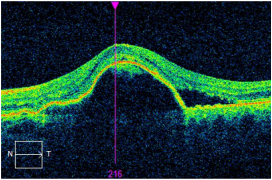
Optical Coherence
|
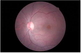
Retinal
|
Office Hours:
Monday/Wednesday/Thursday/Friday 8:30am - 4:30pm
Tuesday 8:30am - 7:00pm
Tuesday 8:30am - 7:00pm